Kelly Avila is a Post Doctoral Researcher working with CucCAP Plant Pathologist Dr. Lina Quesada-Ocampo in the NC State Vegetable Pathology Lab. Kelly’s hometown is Bogota, Colombia and she chose to work in the Quasada lab because of the focus on Extension, integrated disease management, and genomics. Her primary research project is Black rot of sweetpotato and vectors in post-harvest.
Introduce yourself—your background, where you are now, and your current research focus.
I have a major in Biology and a master’s degree in Environmental Science. I did my Ph.D. at a prestigious public university in Colombia, Universidad Nacional de Colombia. I also had the opportunity to do my thesis in a research center named Cenipalma (Research Center in Oil Palm). I have been at NC State since June 2023 working as a Postdoc. In these few month I’ve felt so motivated and excited while learning about this important disease
Why did you choose to work with plant pathology and crop production?
I chose the Quesada Lab for the extension focus.
What do you hope to accomplish during your time working on the CucCAP grant, and what do you most look forward to in this position?
I hope to help improve sweet potato crop management at the post-harvest level. I would like to design a molecular technique for rapid detection of the fungus (C. fimbriata), for example, a LAMP PCR.
What is your favorite plant disease?
Phytophthora pod rot of Cacao
Do you have any social media handles that you want included?
LinkdIn and X (formerly Twitter)
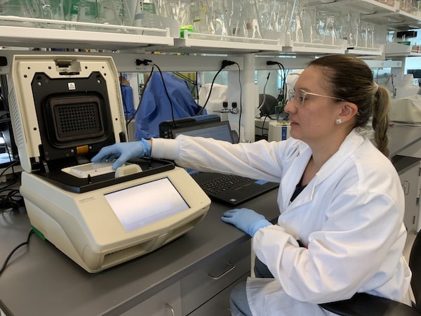
Image 1. Kelly uses a thermocycler for doing qPCR, detecting specific targets of Ceratocystis fimbriata. Nowadays one of the most important techniques in molecular biology studies is polymerase chain reaction (PCR). The method is used to make millions of copies of a specific DNA sequence. With many variants, one of them is qPCR (real-time PCR), which is currently being used in different labs for different research purposes. The equipment in the image is named thermocycler (where traditionally a PCR is run), and this specifically, has an interesting characteristic which is that it reads fluorescent, using specific fluorescent probes you can detect your target DBNA region with this fluorescence to make specific analysis about your biological model.
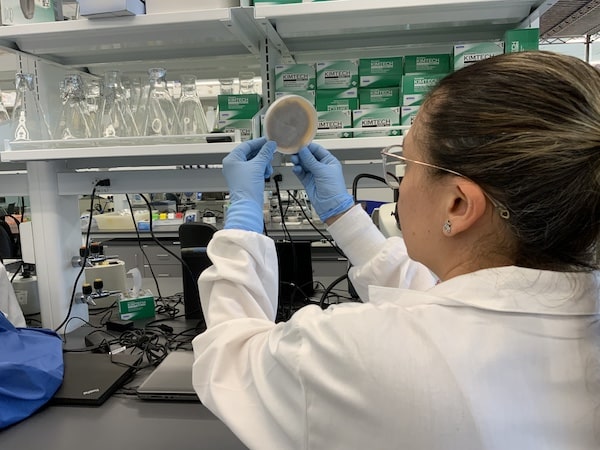
Image 2. Kelly is examining an Agar-plate with Ceratocystis fimbriata. For studying the plant-pathogen infection process, the researcher needs to grow the pathogen of interest under controlled conditions. In this image, there is a petri dish with a growing medium for fungus. In this way, the scientist can see some structures that are related to an organism.
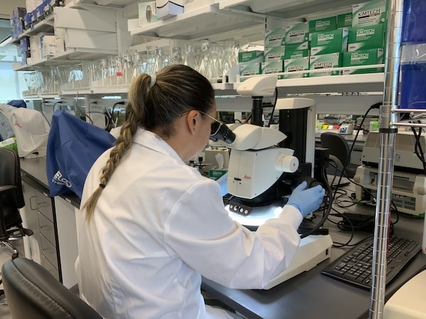
Image 3. Kelly analyzes fungus structures on an agar-plate with a stereoscope. There is a world beyond our eyes that needs to be described, analyzed and counted. Millions of microscopic species exist around us including fungus. It is possible to see some fungus structures with the naked eye, but to describe specific details, the researcher needs to use a microscope or stereoscope.
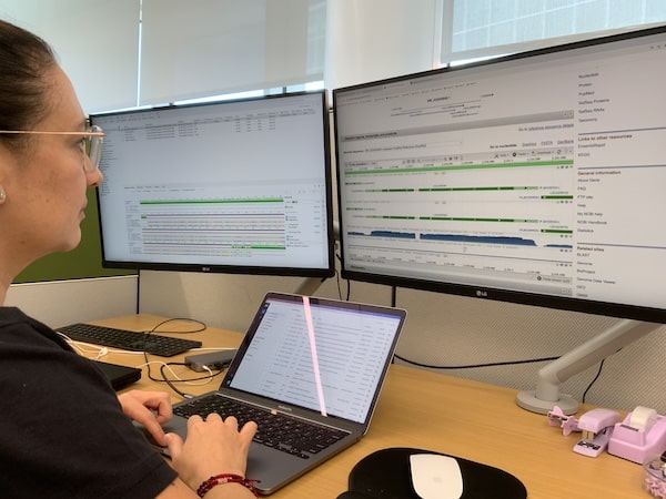
Image 4. Kelly uses online resources for Sequence analysis. In addition to wet-lab duties, bioinformatics is an important discipline that is now being used around the world. Since the growth in availability of sequencing facilities and techniques, labs can access or create genomes for studying biological models with different approaches (omics).
